
(PDF) Upper eyelid and medial canthus reconstructive surgery after
Histiocytosis is a condition with white blood cells that form tumors (histiocytomas) in tissues and organs, like the skin, bones, spleen, liver, lungs, and lymph nodes. There are two types of histiocytomas in dogs, canine cutaneous histiocytoma and malignant histiocytoma. Histiocytes are leukocytes or white blood cells that occur in the tissues.
Histiocytomas in House Pets Lazy Paw Vet Library
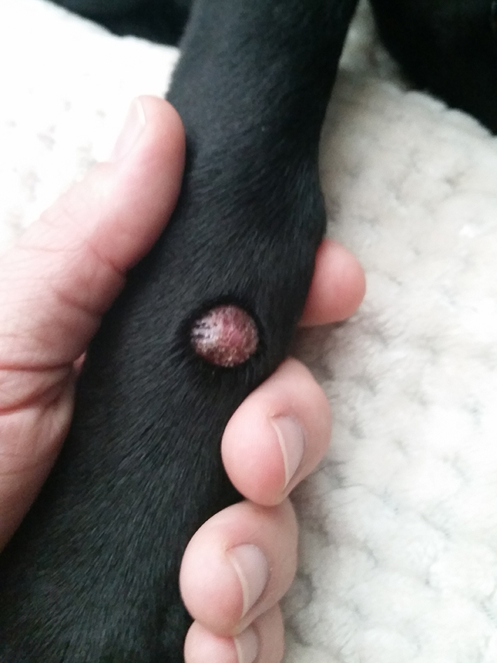
Canine Histiocytoma is a benign, pedunculated, or nodular neoplasm that arises from monocyte-macrophage cells in the skin. They are usually firm and well-circumscribed but sometimes, on palpation feel soft. The overlying hyperpigmented skin is keratotic or shiny and when the tumor is pinched, there is a depression on the surface.

Histiocytoma (dog) Wikipedia
Causes of Histiocytoma in Dogs. The cause of the condition is due to a dog's immune system. Specifically, the growths are caused by the Langerhans cell. Generally, younger dogs under the age of.

5 Canine Histiocytoma Home Treatment
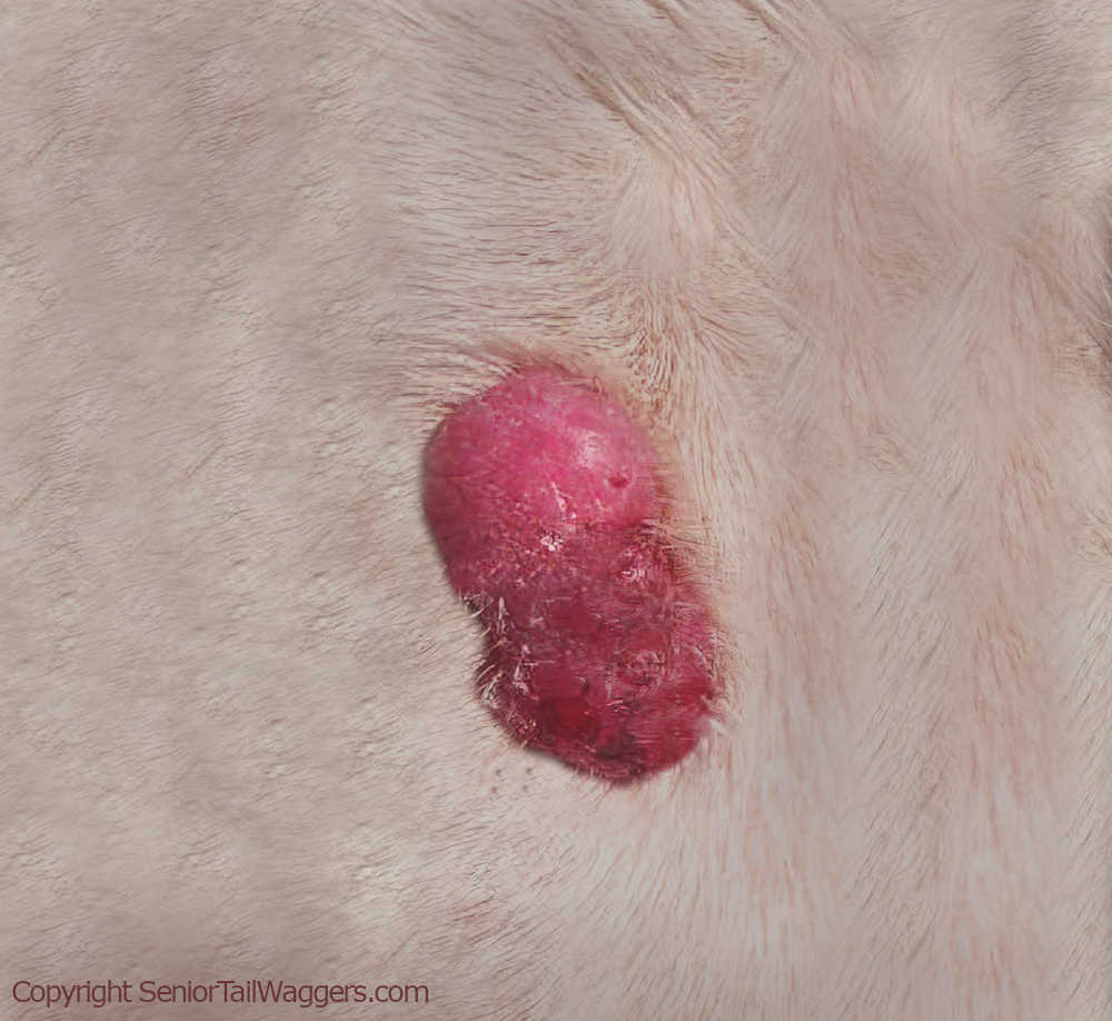
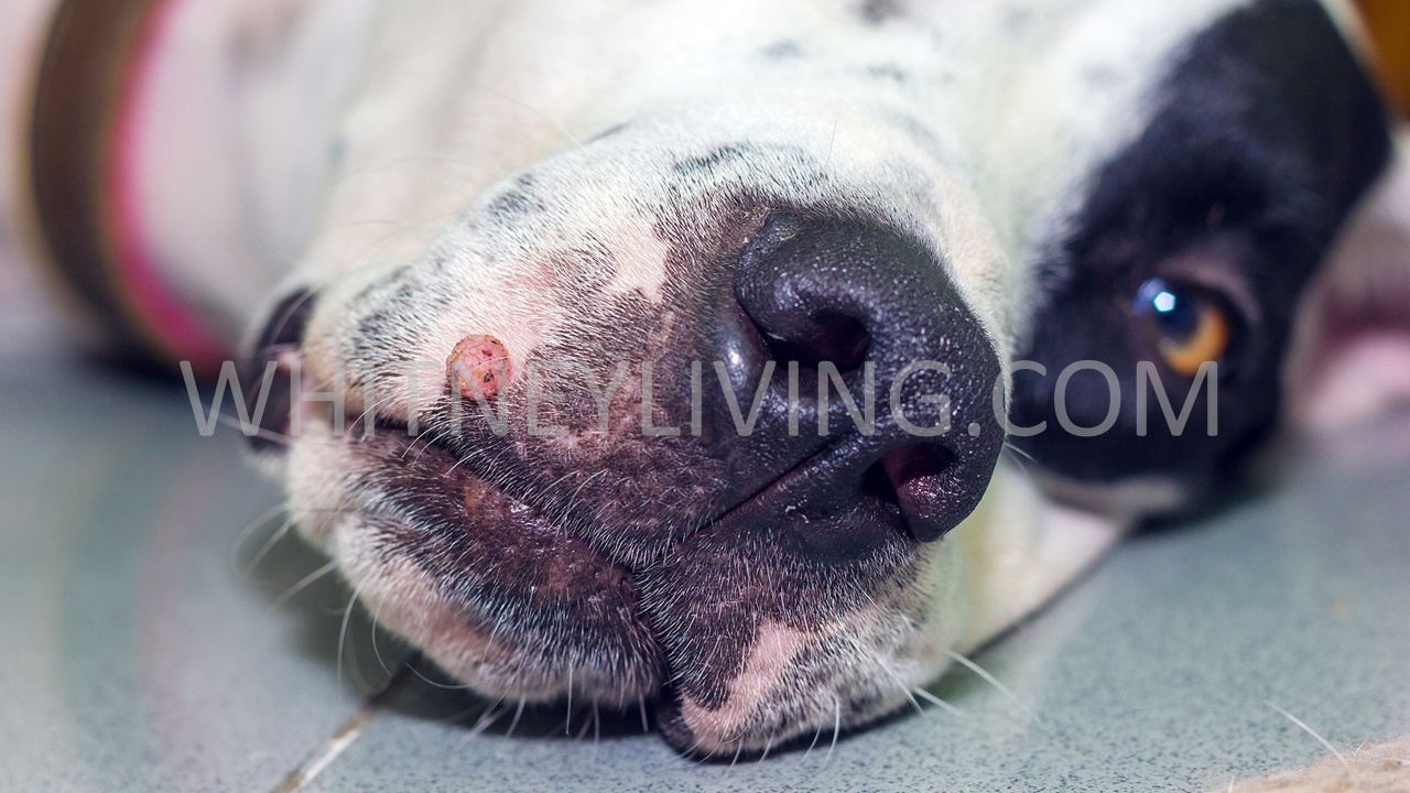
Symptoms & Signs. Histiocytomas are usually raised, red, hairless growths that occur on the head, neck, trunk, or front legs. Histiocytomas usually occur in dogs under two years of age, but they have been known to occur in older dogs as well. Older dogs may develop histiocytomas anywhere on the body.

Pin on dog care
Treatment Costs of Histicytoma In Dogs. The cost of a Histiocytoma removal and biopsy can range from $300 - $1,200 depending on location and provider. This may or may not include the cost for your initial visit and may increase if your pet has other conditions or is geriatric.

7 Clinical Signs of Histiocytoma in Dogs Dogs, Mast cell tumor dogs
The histiocytoma is an unsightly but benign skin tumor that tends to arise on the skin of young dogs. While young dogs (under three years of age) are more likely to get these (especially on the face and extremities), histiocytoma in dogs can happen at any age in just about any location.
:max_bytes(150000):strip_icc()/what-is-a-histiocytoma-3384906_FINAL-5bacfa95c9e77c0025469c05.png)
How to Treat Histiocytomas in Dogs
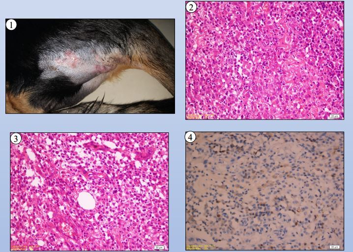
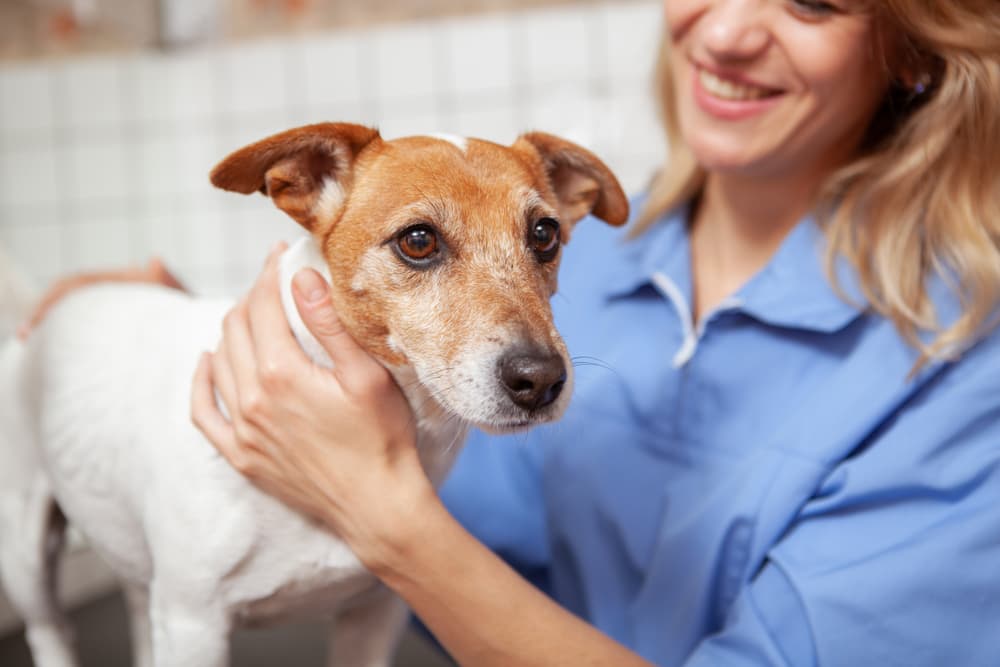
How Vets Diagnose Histiocytomas in Dogs. Often, veterinarians make an initial diagnosis of histiocytoma in dogs based on: The appearance of the growth. The location of the growth. The dog's breed and age. A definitive diagnosis requires microscopic testing, typically through a needle biopsy of the growth. Treatment for Histiocytomas in Dogs

Button tumor (histiocytoma on labrador retriever Pets, People and, Life
Histiocytomas are a type of benign skin mass or "tumor," meaning they are non-cancerous or not malignant. Read on to learn more about what causes them, what they look like, and how they're treated. Causes of Histiocytomas in Dogs What do histiocytomas look like? How are histiocytomas diagnosed in dogs?
The Use Of Lomustine Treatment In A Dog With Multiple Cutaneous
Treatment Prognosis Prevention Histiocytomas look scary but they are not dangerous. Raised, red, and sometimes ulcerated, these benign growths are not usually painful or itchy for dogs. Surgical treatment is only recommended if the bump grows large enough to bother the dog or the owner.
Histiocytomas in Dogs Pictures & Veterinarian Advice
Signs of histiocytomas are much what you'd expect: a red, raised, rounded growth protruding from the skin. They tend to be hairless or sparsely haired. You may first notice them while petting your dog, when they may be smaller and still hidden in the haircoat. However, histiocytomas can grow to be multiple centimeters in size.
5 Histiocytoma Dog Home Treatment And Cure
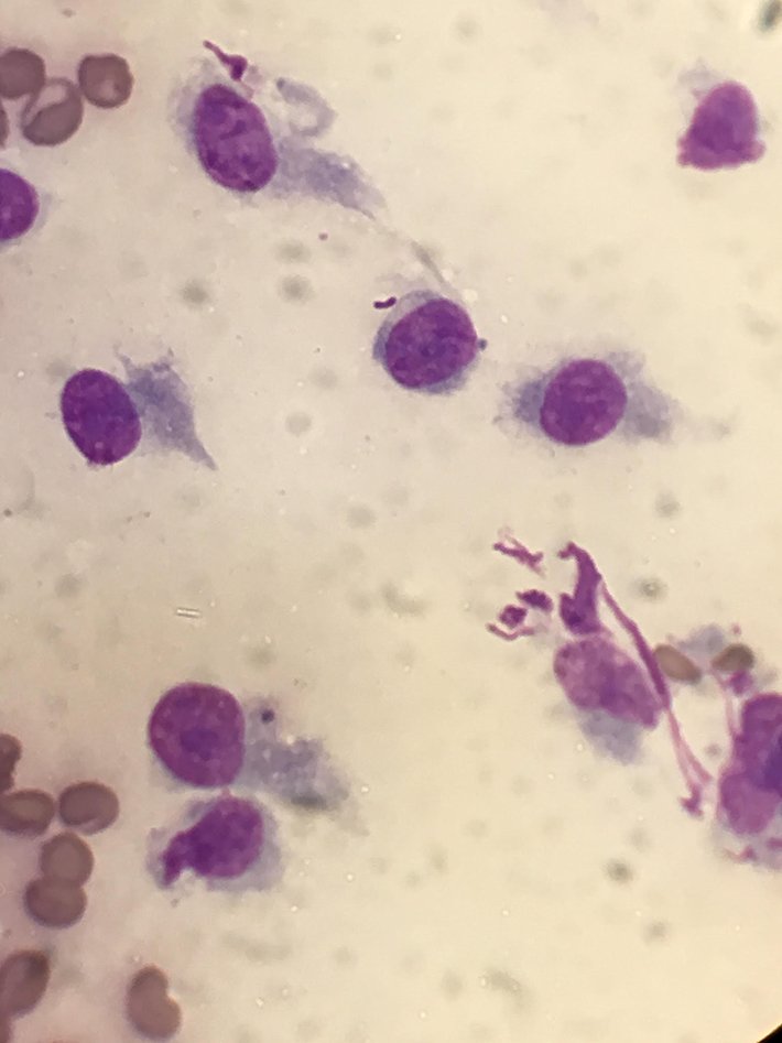
Diagnosis of Histiocytoma in Dogs Diagnosis depends on getting a tissue sample to be able to examine it under a microscope. This is a simple procedure that can be carried out without taking much time. Your veterinary caregiver will use a needle or a punch biopsy to take a bit of tissue for examination.
Canine Histiocytoma Complex I Love Veterinary Blog for
A cutaneous histiocytoma (not to be confused with histiocytosis) is a common, harmless (benign) tumor of Langerhans cells. In the tumor's early stages, over the first one to four weeks, the cells grow rapidly. During this rapid growth, they often ulcerate and may become infected. Later, they may regress spontaneously.
Histiocytomas in House Pets Lazy Paw Vet Library
A histiocytoma is a benign tumor that usually occurs in younger dogs. They are notorious in Boxers. They are generally rapid-growing, hairless, red spots found on your dogs' skin. They can regress and go away on their own but are also commonly removed via surgery.

Histiocytoma in Dogs Symptoms, Treatment and Prevention Health info
A Histiocytoma is a growth that develops on the surface of a dog's skin. Histiocytomas are benign, non-cancerous nodules, commonly known as round cell tumors. Histiocytoma can occur in any breed of dog but boxers, bulldogs, and flat-coated retrievers are the more commonly affected breeds. Histiocytomas are not contagious and they tend to be.
Histiocytoma in Dogs Great Pet Care
Histiocytoma is a tumor that originates from histiocytes, a group of lymphoid cells that are an essential part of the dog's immune system. Cutaneous histiocytoma is a very common benign tumor in dogs. One report from the United Kingdom claims that histiocytoma is the most common single tumor type in dogs [1].

9040d1267310196 histiocytoma dsc05053 Dog skin problem, Dog skin, Pet
Key Takeaways You generally do not need to treat histiocytoma in dogs. Most will regress and disappear on their own, usually in two or three months. A histiocytoma that becomes infected or is otherwise irritating may need to be surgically removed rather than waiting for it to disappear.